
Digital X-Rays
Digital X-Rays
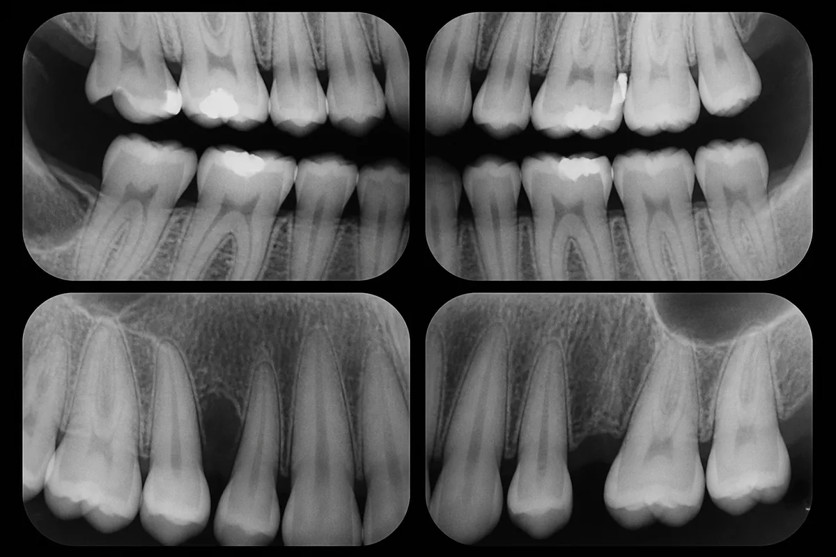
Digital X-rays use small sensors instead of traditional film to capture images of your teeth, roots, and supporting bone. The image appears on a screen within seconds, allowing your dentist to review it right away as part of your exam. Because digital sensors are more sensitive than film, they typically require less radiation to produce a clear picture, and the images can be enlarged or adjusted for a closer look at areas of concern.
How Digital X-Rays Fit Into Your Visit
- Cavity Detection — Decay between teeth or beneath old fillings often isn’t visible during a routine look in the mouth.
- Bone and Root Health — Images show bone levels and root structure, which is useful for monitoring gum health and planning treatment.
- Treatment Planning — Digital radiographs support planning for fillings, crowns, extractions, and other restorative work.
Part of Our Digital Approach
Digital X-rays are one part of the imaging available at our Chicago office. For a broader view of the jaw, teeth, and sinuses, our Panoramic X-Ray captures a single wide image, while 3D Cone Beam CT Imaging is available when a more detailed view of bone density and nerve paths is needed, such as for implant planning. All images are stored as part of our Fully Digital Records, so your imaging history is accessible across our offices whenever you need care.
This content is for general information only and isn't dental or medical advice. Talk with your dentist or orthodontist about your own situation. If you have a medical emergency, call 911.