
3D Cone Beam CT Imaging
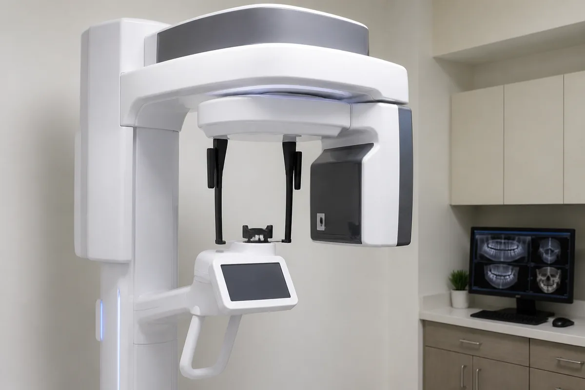
3D Cone Beam CT Imaging
3D Cone Beam CT Imaging gives our dentists a detailed, three-dimensional view of your teeth, jawbone, nerve pathways, and surrounding soft tissue in a single scan. It's a tool we rely on when treatment planning needs more information than a flat, two-dimensional picture can provide, particularly for implants, oral surgery, and other cases where precision matters.
What 3D Cone Beam CT Imaging is
Cone Beam Computed Tomography, or CBCT, is a type of dental x-ray that captures a full three-dimensional volume of your mouth, jaw, and facial structures instead of a single flat image. During the scan, an arm rotates around your head, taking a series of images from different angles. A computer then combines these into one detailed 3D model that can be viewed and measured from any direction.
This is a meaningful step up from traditional dental x-rays or panoramic images, which show teeth and bone in only two dimensions and can hide overlapping structures. With cone beam imaging, bone density, root shape, nerve canal position, sinus location, and soft tissue can all be seen clearly in relation to one another, which is especially useful before any procedure that involves working near a nerve, sinus, or existing tooth root.
How we use it at 1st Family Dental
On-site 3D Cone Beam CT Imaging lets our clinicians see the exact position of bone, nerves, and tissue before recommending or performing surgical treatment, so a plan can be built around your actual anatomy rather than an estimate. This works hand in hand with our guided implant surgery, where a surgical guide is fabricated directly from the scan data so each implant can be placed exactly where it was planned.
Team members whose training includes CT-based planning apply this imaging in their own areas of focus. Dr. John Hur, an Oral and Maxillofacial Surgeon, uses CT-guided skeletal mapping as part of implant and corrective jaw surgery. Dr. Najeeb Abufarha's continuing education includes 3D imaging as part of digital treatment planning for implant placement and bone grafting. Orthodontists Dr. Budi Kusnoto and Dr. Sameer Al-Obaidi have training in 3D imaging as it applies to orthodontic diagnosis and treatment planning, including cases involving temporary anchorage devices. The resulting images are stored as part of our fully digital records, so they can be reviewed alongside panoramic x-rays and shared securely if your care involves more than one provider.
What it means for you
A single cone beam scan can reduce the need for multiple separate x-rays, since it captures bone, teeth, nerves, and soft tissue together. That makes it easier for your dentist to explain what they're seeing and why a particular treatment approach is recommended, using the same 3D images you're looking at.
- Clarity — structures that overlap on a flat x-ray are separated out in three dimensions.
- Precision — surgical guides and implant plans can be built from your actual bone anatomy.
- Efficiency — one scan can replace the need for several individual images.
- Better conversations — you can see the same images your dentist is reviewing when discussing options.
What to expect during your visit
If your dentist recommends a cone beam scan, it's typically done as part of a treatment planning appointment, such as before implant placement, oral surgery, or a complex orthodontic case. You'll be asked to remove metal jewelry or eyewear and may wear a protective apron, similar to a standard dental x-ray.
During the scan, you'll stand or sit still while the imaging arm rotates around your head. There's no injection and nothing touches your teeth or gums. The rotation itself generally takes well under a minute, though you may be positioned briefly beforehand to make sure the area of interest is captured. Once complete, the images are processed digitally and can usually be reviewed with your dentist that same visit.
Is 3D Cone Beam CT Imaging safe?
Cone beam scanning uses x-rays, the same type of energy used in standard dental imaging, and is a widely used diagnostic tool in dentistry. Your dentist will determine whether a scan is appropriate for your specific situation before recommending one.
Will the scan be uncomfortable?
The scan itself does not involve any contact with your teeth or gums, and there is no injection involved. You'll simply hold still while the imaging arm moves around your head for a short period of time.
How long does the imaging appointment take?
The scan itself is quick, generally under a minute of actual rotation, though the full appointment may include positioning and a short review of the images with your dentist.
Who typically needs this type of imaging?
Cone beam imaging is commonly used before procedures where precise anatomy matters, such as dental implant placement, oral surgery, bone grafting, or certain orthodontic and TMJ evaluations. Your dentist will let you know if your treatment plan calls for this type of scan.
If you're planning treatment that may benefit from a detailed 3D view of your teeth and jaw, reach out to 1st Family Dental to ask whether Cone Beam CT Imaging is right for your care.
This content is for general information only and isn't dental or medical advice. Talk with your dentist or orthodontist about your own situation. If you have a medical emergency, call 911.